Breast cancer happens to be one of the most prevalent types of cancers that affect women around the world. Early diagnosis increases the possibility of treatment. As such, it is important to understand how important breast health is and to learn when to screen for any problems. Nevertheless, there are people who are uncertain about the screening procedure, the use of mammogram and other tests that they should consider.
A mammogram is a test that is done to find breast abnormalities, which may include cancers even before any signs become visible. It is important to understand how mammography works, the benefits that are associated with mammogram and when professional advice is required.
According to WHO, early diagnosis and treatment of breast cancer is important to improving breast cancer outcomes.
What Is a Mammogram for Breast Cancer?
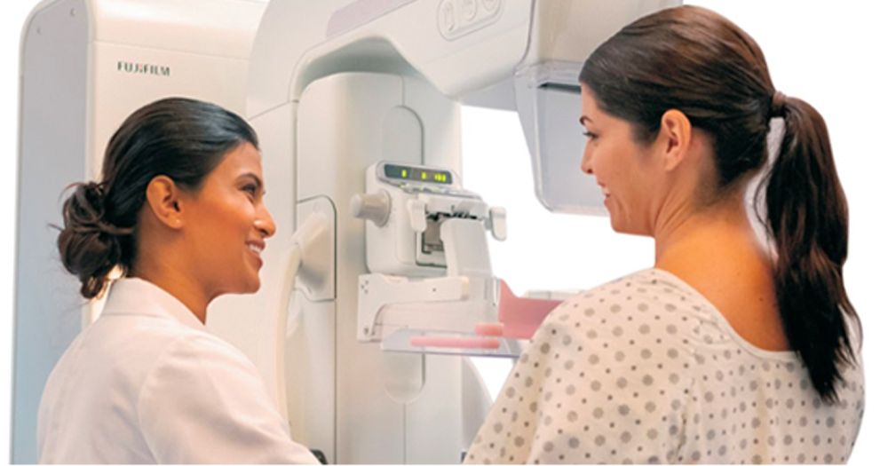
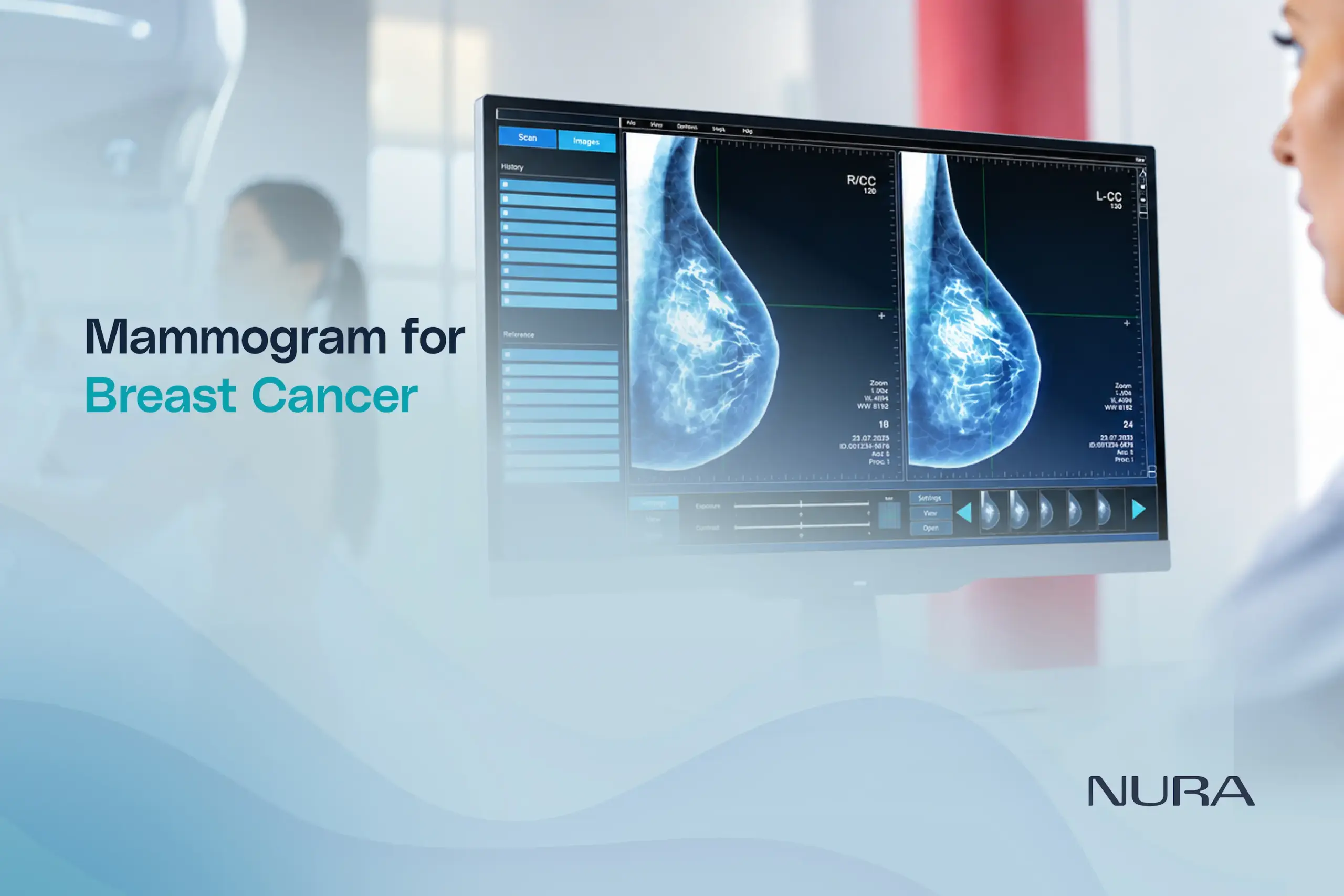
A mammogram refers to an X-ray picture of the breast created through the use of a special imaging device. The mammogram of breast cancer assists medical professionals in detecting abnormalities in the breast tissue that cannot be detected by means of clinical examinations only.
Mammography involves the use of low-dose X-rays to produce an image of the breast. Abnormalities include small lumps, tissue abnormalities and microcalcifications, which are very small deposits of calcium in the breast.
It is important to understand that a mammogram cannot confirm a cancer diagnosis on its own. If a suspicious area is detected, additional tests, such as a breast ultrasound or biopsy, may be recommended to determine the cause. You can also read our blog on how digital mammography helps detect breast cancer early to learn more.
Why Is Mammography Important for Breast Cancer Screening?
One of the main benefits of mammography is its ability to detect certain breast cancers before symptoms develop. Early detection may allow treatment to begin when the disease is at a more treatable stage.
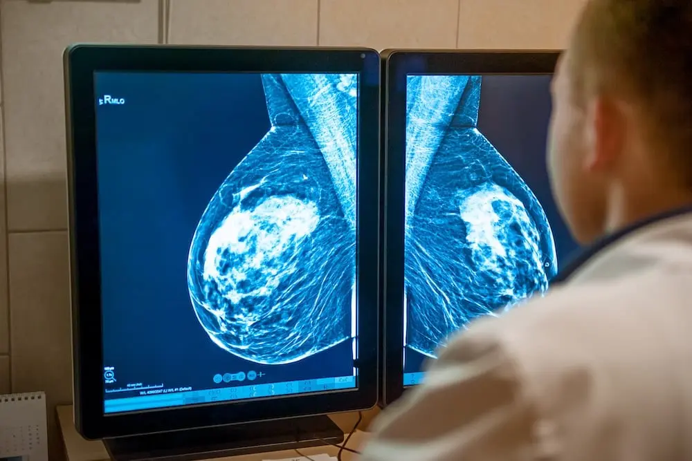
Mammography can also help identify abnormalities that require further assessment, supporting timely clinical evaluation. Nevertheless, not every abnormality is cancer, and not every breast cancer will be detected by a mammogram.
Breast screening is therefore one component of a broader approach to breast health that includes awareness of symptoms, assessment of individual risk, and appropriate follow-up care.
Types of Mammography: Screening vs Diagnostic Mammography
Although both procedures use breast X-ray imaging, screening mammography and diagnostic mammography serve different purposes.
1. Screening Mammogram
The screening mammogram is carried out in individuals without breast-related symptoms as a part of a proper breast cancer screening program.
This test seeks to find any suspicious changes that occur even before they can be felt. In certain cases, depending on the breast screening program and personal situations, one might require a mammogram at regular intervals.
The right age to begin and interval are determined by factors like age, personal history, family history, among others.
2. Diagnostic Mammogram
The diagnostic mammogram is used whenever there is a need to evaluate any symptom of the breast or when a screening mammogram reveals any abnormality.
This procedure might entail some more X-ray shots of a specific part of the breast. A radiologist will examine the pictures to assess whether any other investigations like breast ultrasound or biopsy are needed.
Key difference between the screening mammogram and diagnostic mammogram is that, the screening helps to find out any signs of illness in people who have no symptoms at all, while the diagnostic mammogram investigates any specific problem or abnormality.
When Is a Mammogram Needed?
There is no single screening schedule that is appropriate for everyone. The decision to undergo a mammogram should consider a person’s age, breast cancer risk, medical history, and symptoms.
Based on Age
Age is one factor that can contribute to the increased risk of breast cancer. But guidelines regarding the starting age for regular mammogram screenings can vary from country to country and among professional bodies.
For instance, the United States Preventive Services Task Force advises women ages 40-74 at average risk to have biennial screening mammograms. Different groups provide varying suggestions about screening frequency.
Nevertheless, these guidelines must not necessarily apply to everyone without exception. Each person is encouraged to consult a healthcare professional for an ideal screening schedule.
Family History and Increased Risk
Individuals who have a family history of breast cancer, inherited gene mutations like BRCA1 and BRCA2, or a history of breast cancer themselves might need a personalised approach to screening.
Exposure to radiation to the chest at a young age can also put someone at risk for developing breast cancer.
It will be up to the healthcare provider to determine how often and what kinds of imaging tests, such as breast MRI, are needed. Breast cancer screening must be done according to a proper risk assessment, not only because of family history.
When Breast Symptoms Appear
Any individual who recognizes that there is a new breast lump, changes to nipples, any abnormal discharge from breasts, or any changes in the skin of the breast needs to seek a medical examination without delay.
It is up to the doctor’s discretion to perform a diagnostic mammogram, an ultrasound scan, or other tests. People need not wait for the next routine breast screening session to be done, when such changes occur.
Having a normal breast screen mammogram does not mean one cannot have breast cancer.
What Happens During a Mammogram?
Understanding the procedure can help reduce uncertainty and anxiety before an appointment.
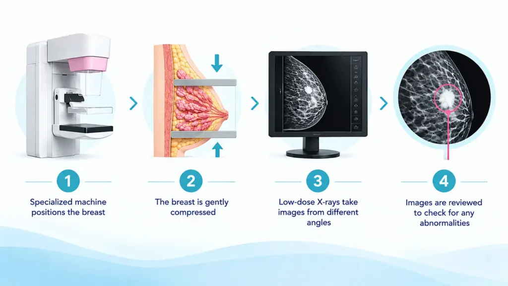
Step 1: Preparation
The imaging team might inquire about prior breast imaging, breast symptoms, medical history, and pregnancy. It is advisable to carry any old mammography reports/images if possible.
It should be avoided to use any deodorant, talcum powder, or lotion on the breasts or armpit area on the day of the test, if advised by the imaging facility, since such items may be mistaken for markings in the images.
Step 2: Positioning
The radiographer positions the breast on a specialised imaging platform. A second plate gently compresses the breast to spread the tissue and obtain clear images while using a low radiation dose.
Step 3: Taking the Images
The breast undergoes compression for a short period as X-ray pictures are taken. There might be some discomfort from pressure, but this discomfort varies from person to person.
The process is repeated to obtain the required views of both breasts or the area being investigated.
Step 4: Reviewing the Results
A radiologist interprets the images and prepares a report. If the findings are reassuring, the person may be advised to follow the recommended screening schedule. If an abnormality is identified, additional imaging or a biopsy may be required.
Mammogram vs Breast Ultrasound: What Is the Difference?
Mammography and breast ultrasounds are two separate imaging methods, and both play unique roles when it comes to the evaluation of the breast.
Mammograms use low doses of X-rays for detecting abnormal areas, which sometimes can be hard to detect by touch. Ultrasound scans use sound waves to create an image of the breast and differentiate cystic and solid breast masses.
Ultrasound scans may be required to examine a lump, to clarify the findings of a mammogram, or to evaluate some abnormalities in the breast.
However, breast ultrasound is not a universal replacement for mammography in routine breast cancer screening. The choice of imaging depends on age, symptoms, breast density, risk factors, and clinical findings.
Are Mammograms Safe? Understanding Their Limitations
Mammography is done using low doses of ionizing radiation. In cases where mammography screening is recommended to individuals, the benefits of the procedure need to be balanced with its disadvantages. These include:
- False-positive results: An image may appear suspicious even when cancer is not present, leading to additional tests.
- False-negative results: Some breast cancers may not be visible on a mammogram.
- Additional investigations: An abnormal finding may require further imaging or a biopsy.
- Discomfort: Temporary pressure or tenderness may occur during breast compression.
- Overdiagnosis: Screening can sometimes identify cancers that would not have caused symptoms or affected a person’s health during their lifetime.
Can Breast Cancer Be Detected Without a Mammogram?
Being familiar with the usual appearance and feel of your breasts can help you recognise changes that require medical attention.
Warning signs may include:
- A new lump or thickening in the breast or underarm.
- Changes in breast size, shape, or appearance.
- Dimpling, puckering, or unusual changes in the breast skin.
- A newly inverted nipple or persistent nipple changes.
- Unusual nipple discharge, particularly if bloody.
These signs are not always an indication of cancer, but must be evaluated by a health care professional. Breast cancers sometimes occur in the time period between mammograms, hence the importance of being aware of symptoms even after a normal mammogram.
Make Breast Health Part of Preventive Healthcare
Preventive healthcare comprises knowledge about one’s risk factors, undergoing proper screenings, and consulting with a doctor if any symptoms occur. In the case of breast cancer, mammograms can play a crucial role in early detection as long as they are conducted properly. However, each individual may need a different type of screening, which will be determined after a discussion with a competent healthcare professional. At NURA, the focus on preventive health screening reflects the importance of informed health decisions and early identification of potential health concerns. People who have been thinking about breast imaging need to address the healthcare professional to get more information about the available screening methods.
Preventive health checkups can also help women take a more proactive approach to their overall well-being. NURA offers a women’s health checkup in Chennai, Bengaluru, Gurugram, Mumbai, Hyderabad, and Calicut that includes screening for breast and cervical cancer, along with assessments for various lifestyle-related health risks. Choosing an appropriate screening package based on age, personal medical history and individual risk factors can help women make informed decisions about their health.


For those looking for a broader assessment of their health, a master health checkup in Chennai, Bengaluru, Gurugram, Mumbai, Hyderabad, and Calicut at NURA provides a comprehensive preventive screening experience covering multiple body systems. The screening combines Ai-assisted imaging, laboratory tests and doctor consultation, helping individuals understand potential health risks and identify areas that may require further medical attention.